Structural connections between the language cortices
Different parts of the brain are highly interconnected with other parts by fiber tracts. In the 19th century these fiber tracts were identified in vitro. Nowadays, diffusion tensor imaging (DTI) allows to identify structural connections between different brain regions in the human in vivo (e.g. Behrens et al., 2003; Johansen-Berg et al., 2004). For a recent tractography atlas representing the major fiber connections based on this method see Catani & de Schotten (Catani & de Schotten, 2008). Note, however, that with this approach the directionality of the connection cannot be determined. An important fiber pathway connecting two language regions, namely Broca’s area and the temporal cortex (Wernicke’s area) is the arcuate fasciculus (AF) and the superior longitudinal fasciculus (SLF). Concerning the connections between the language-relevant regions, the literature generally discusses two pathways, a dorsal (AF/SLF) and a ventral pathway.
‘Dual stream models’ (Hickok & Poeppel, 2004; 2007; Rauschecker & Scott, 2009) assume that the ventral pathway supports sound-to-meaning mapping whereas the dorsal pathway connecting the posterior dorsal-most aspect of the temporal lobe and the posterior frontal lobe supports auditory-motor integration (Hickok & Poeppel, 2007). Recently, there has been a debate with respect to the particular functions of different pathways from the temporal cortex to other parts of the brain as well as with respect to their endpoints in the other brain regions (see Friederici, 2009a; 2009b; Weiller et al., 2009) (see Fig. 4). For language processing Saur et al. (2008, 2010) proposed the ventral pathway connecting the temporal cortex with the pars orbitalis (BA 47) and triangularis (BA 45) via the extreme capsule fiber system (ECFS) supports sound-to-meaning mapping, whereas the dorsal pathway connecting the temporal lobe to the premotor cortex and continuing to the pars opercularis (BA 44) supports sensory-motor mapping of sound-to-articulation. In their studies Saur and colleagues used a deterministic fiber tracking approach in which the two end points of the connection were predefined on the basis of functional data. Friederici et al. (2006a) used a probabilistic fiber tracking approach in which only one end of the connection is defined as a seed point. Two seed points in the IFG based on two functionally different activations revealed a dorsal pathway going from pars opercularis (BA 44) to the posterior temporal cortex via the arcuate fascicle/superior longitudinal fascicle (AF/SLF), and a ventral pathway from the frontal operculum (FOP) via the uncinate fascicle (UF) to the anterior temporal cortex. The dorsal pathway was interpreted to support the processing non-adjacent elements in syntactically complex sentences and the ventral pathway to support combinations of adjacent elements in a sequence.
These findings suggest that there are two ventral pathways and two dorsal pathways connecting the frontal to the temporal cortex involved in language processing. Ventral Pathway I goes from BA 45 via the ECFS to the temporal cortex and Ventral Pathway II goes from the FOP via the uncinate fascicle (UF). Dorsal Pathway I goes from the temporal cortex to the pre-motor cortex and Dorsal Pathway II goes from the temporal cortex to BA 44. Dorsal Pathway I may mainly support sound-to-motor mapping whereas Dorsal Pathway II may rather support higher-level language processes (for a recent debate, see Friederici, 2009a; 2009b; Weiller et al., 2009).
To summarize, there are multiple long-range structural connections between the language-relevant regions in the frontal and temporal cortices: two dorsal pathways and possibly two parallel ventral pathways.
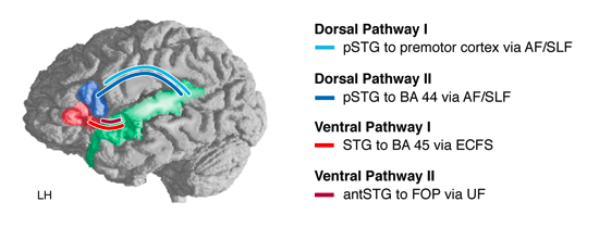
Figure 4: Structural connectivities between the language cortices
Schematic view of two dorsal pathways and two ventral pathways. Dorsal pathway I connects the superior temporal gyrus (STG) to the premotor cortex via the arcuate fascile (AF) and the superior longitudinal fascicle (SLF). Dorsal pathway II connects the STG to BA 44 via the AF/SLF. Ventral pathway I connects BA 45 and the temporal cortex via the extreme capsule fiber system (ECFS). Ventral pathway II connects the frontal operculum (FOP) and the anterior temporal STG/STS via the uncinate fascile (UF). Figure legend is taken from Friederici, 2011 (Physiological Reviews, 91(4), 1357-1392).