Parcellation of the language cortex: Receptorarchitectonic analysis
An entirely novel approach bases the parcellation of the cortex on a receptorarchitectonic analysis which considers the distribution of different types of neurotransmitter systems (e.g. Noradrenalin, Acetylcholin, GABA, Glutamat, Serotonin, Dopamin, Adenosin etc.) in the cortex (Amunts et al., 2010, Zilles & Amunts, 2009).
Frontal cortex
A receptorarchitectonic analysis of the left prefrontal cortex reveals, a more fine grained parcellation than the cytoarchitectonic analysis. Area 45 can be subdivided into two parts, a more anterior area 45a bordering BA 47 and a more posterior area 45p bordering BA 44 (Amunts et al., 2010) (see Fig. 3). Area 44 can also be receptorarchitectonically subdivided two parts a dorsal (44d) and a ventral area (44v). These subdivisions may be of particular functional relevance for language, since different language studies have allocated different functions, such as semantics and syntax, to different locations within area 45, and area 44. These may now be assigned to different subregions within 45 (45a vs 45p) and 44 (44d vs 44v).
Temporal cortex
Receptorarchitectonic analyses of the temporal cortex are not yet available, but studies are ongoing.
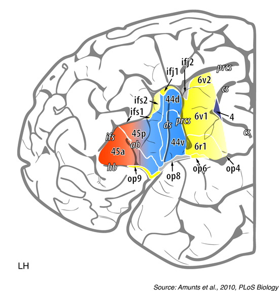
Figure 3: Receptorarchitectonic parcellation of the left posterior prefrontal cortex
Extent of delineated areas projected to the lateral surface of an individual post mortem brain. The following receptor binding sites were studied by Amunts et al. (2010) for the prefrontal cortex: glutamatergic AMPA and kainite receptors, GABAergic GABAA receptors, cholinergic muscarinic M1 and M2 receptors, and noradrenergic receptors. The color coding indicates cytoarchitectonically defined borders. The borders between 44 d (dorsal) and 44 v (ventral), for example, was differentiated mainly by α 1 and muscarin M2 receptors. Area 45 can be subdivided receptorarchitectonically into an anterior (45a) and a posterior (45p) part. Area 6 can be subdivided into three subparts. Abbreviations: op = operculum (numbering indicates different subparts), ifs = inferior frontal sulcus, ifj = inferior frontal junction, prcs = precentral sulcus, cs = central sulcus. Figure legend is taken from Friederici, 2011 (Physiological Reviews, The Brain Basis of Language Processing: From Structure to Function).