Parcellation of the language cortex: Cytoarchitectonic analysis
Korbian Brodmann (Brodmann, 1909) was the first neuroanatomist to provide a cytoarchitectonic description of the human cortex in the left hemisphere, analyzing one single brain. A more recent approach provides a semi-anatomically objective cytoarchitectonic analysis by calculating the density of different types of neurons in the different cortical layers in a number of brains leading to a more generalized map (Amunts et al., 1999; Amunts & Zilles, 2001; Link). This analysis allows to segregate different cortical regions into subregions.
Frontal cortex
In the frontal cortex of the left hemisphere the cytoarchitectonic approach proposes a subdivision of Broca’s area. Such a subdivision appears to be of particular importance since the region of Broca’s area has often been discussed as supporting different subprocesses during language processing (Bookheimer, 2002; Hagoort, 2005; Poldrack et al., 1999). Broca’s area is defined to consist of at least two parts, i.e. the cytoarchitectonically defined Brodmann area (BA) 44, the pars opercularis and BA 45, the pars triangularis (Amunts et al., 1999; Brodmann, 1909) (see Fig. 2).
Temporal cortex
The cytoarchitectonic description of the auditory and temporal cortices has only been refined recently. In the primary auditory cortex (PAC), classically BA 41, new cytoarchitectonic analyses have revealed three subregions in a medial-to-lateral direction with Te1.0 in the middle, Te1.1 located more medially and Te1.2 located more laterally (Morosan et al., 2001). The classical cytoarchitectonic analysis by Brodmann (1909) defined one region namely BA 22 to cover the posterior two thirds of the lateral convexity of the superior temporal gyrus (STG) (see Fig. 2). More recent cytoarchitectonic analyses have proposed a separation of the dorsal and ventral banks of the STG (:source: Morosan et al., 2005). For language processing the lateral STG proper excluding the dorsal and ventral banks is functionally relevant.
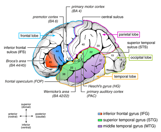
Figure 2: Anatomical and cytoarchitectonic details of the left hemisphere
The different lobes (frontal, temporal, parietal, occipital) are marked by colored borders. Major language relevant gyri (IFG, STG, MTG) are color coded. Numbers indicate language relevant Brodmann Areas (BA) which Brodmann (1909) defined on the basis of cytoarchitectonic characteristics. The coordinate labels superior/inferior indicate the position of the gyrus within a lobe (e.g. superior temporal gyrus) or within a BA (e.g. superior BA 44; the superior/inferior dimension is also labeled dorsal/ventral). The coordinate labels anterior/posterior indicate the position within a gyrus (e.g. anterior superior temporal gyrus; the anterior/posterior dimension is also labeled rostral/caudal). Broca’s area consists of the pars opercularis (BA 44) and the pars triangularis (BA 45). Located anterior to Broca’s area is the pars orbitalis (BA 47). The frontal operculum (FOP) is located ventrally and more medially to BA 44, BA 45. The premotor cortex is located in BA 6. Wernicke’s area is defined as BA 42 and BA 22. The primary auditory cortex (PAC) and Heschl’s gyrus (HG) are located in a lateral to medial orientation. Figure legend is taken from Friederici, 2011 (Physiological Reviews, 91(4), 1357-1392).