Syntactic complexity and experimental demands
Almost all neuroimaging studies on syntactic complexity report activation in the inferior frontal gyrus. The majority found the activation located in Broca’s area, namely BA 44 and BA 45, but some also report BA 47 and BA 6 to be involved. The variation in the localization of the syntactic complexity effect may be due to different task demands or different input conditions used in the various studies.
Evidence that the task demands are at least part of the explanation for the observed variation of localization across the different studies come from a study that directly compared three different tasks, i.e. plausibility judgement, sentence verification and non-word detection in a within-subject design (Caplan, Chen & Waters, 2008). While all three tasks activated BA 44 other brain regions were also activated differing for the three tasks. Thus BA 44 was identified as the core region of syntactic computation.
A second factor that might influence the localization of the syntactic complexity effect is the quality of the input. Recently, it was shown that German object-first sentences versus subject-first sentences showed more activation in BA 44 and in the posterior superior temporal sulcus when presented as normal speech, but the localization of this syntactic complexity effect shifted when the auditory input was degraded (Obleser et al., 2011). In the inferior frontal cortex it shifted from BA 44 to a more superior and posterior region, namely the inferior frontal sulcus, and in the superior temporal cortex it shifted towards the auditory cortex (see Figure 9).
In sum, it appears that the syntax complexity effect can shift its maximum within the IFG, both as a function of task demands and input conditions. This point towards a language processing system, which allocates different subregions in the perisylvian default language network as needed.
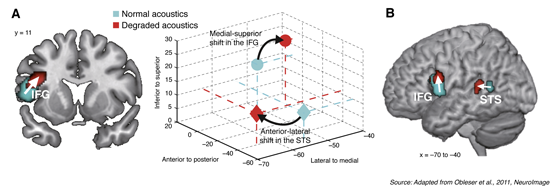
Figure 9: Activation shift for syntax as a function of intelligibility
The panels show activation overlays of three major contrasts: syntactic complexity without acoustic degradation in red and syntactic complexity under acoustic degradation in blue. All activations are thresholded at p (uncorr.) < 0.001. A: Maxima of activation (coronal view) for syntactic complexity without and with acoustic degradation in the left IFG. Activation shifts towards superior BA 44 and inferior frontal sulcus. B: Maxima of activations (lateral view) for syntactic complexity without and with acoustic degradation in the IFG and in the left STS. In the STG activation shifts towards the auditory cortex. Middle part of the figure represents these shifts in MNI coordinate system. Figure legend is taken from Friederici, 2011 (Physiological Reviews, 91(4), 1357-1392).