Brain basis of auditory comprehension
Initial acoustic-phonological processes take place during the first 100 ms after acoustic stimulation crucially involve the primary auditory cortex (PAC) and the planum temporale (PT). From these regions, the information is transferred both to the anterior and the posterior superior temporal gyrus (STG) and sulcus (STS). The left anterior STS is sensitive to the intelligibility of a stimulus. The anterior STG, together with the left frontal operculum (FOP) connected via a ventral pathway (Ventral Pathway II, see Figure 13C) through the uncinate fasciculus (UF), builds the neural network for initial local structure building processes. During a second processing step semantic, grammatical and thematic relations in a sentence are processed in parallel systems, activating separable left-lateralized temporo-frontal networks. The semantic network involves the middle and posterior STG/MTG (sometimes extending into anterior temporal regions) and BA 45 (and BA 47) in the frontal cortex connected via another ventral pathway (Ventral Pathway I, see Fig. 13C) through the extreme capsule fiber system (ECFS). The syntactic network dealing with complex sentence structures involves BA 44 as part of Broca's area in the frontal cortex and the posterior STG/STS connected via a dorsal pathway (Dorsal pathway II, see 13C). Note, that Dorsal Pathway I connecting the temporal cortex to the premotor cortex is supposed to support sensory-to-motor mappings (see Fig. 13C). Syntactic and semantic integration processes take place during a third processing step, possibly under the involvement of the posterior STG/STS and the Basal Ganglia.
The processing of suprasegmental prosodic information is supported by the right hemisphere. The interaction of syntactic information processed in the left hemisphere and prosodic information processed in the right hemisphere is supported by the posterior portion of the corpus callosum, the structure which connects the temporal cortices of the two hemispheres.
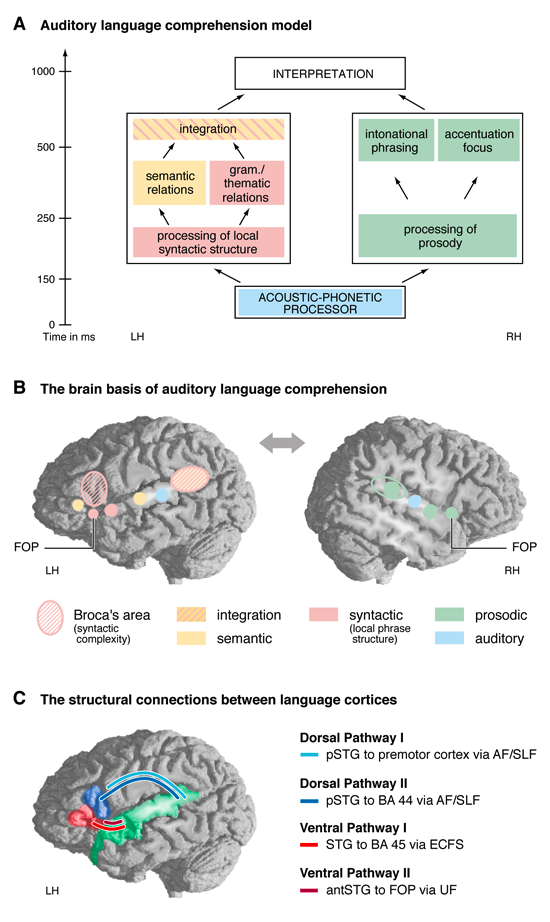
Figure 13: Model of auditory sentence comprehension
A: Auditory language comprehension model. The different subprocesses are indicated by the different boxes. For the exact time course of these subprocesses and their electrophysiological correlates see Friederici, 2011 (Physiological Reviews, 91(4), 1357-1392).
B: The brain basis of auditory language comprehension. Brain regions involved in the different subprocesses labeled in Figure 12A are indicated by activation maxima from prime fMRI studies. Acoustic-phonetic (phonological) processes (blue) involve the primary and secondary auditory cortex located lateral to Heschl’s gyrus in the middle portion of the STG. Processing local syntactic structure (solid pink) involves the anterior STG and the FOP (Friederici et al., 2003). Processing grammatical relations in complex syntax sentences (striped pink) involves Broca’s area and the posterior STG/STS (see Figure 6). Processing semantic relations (yellow) involves the middle and posterior portion of the STG and MTG in the temporal cortex (only the STG activation is indicated in the figure), and also the BA 45/47 in the IFG whenever the task requires controlled processes (Poldrack et al., 1999). Integration of semantic (sem) and syntactic (syn) information (striped pink yellow) involves the posterior portion of the STG/STS. Processing prosodic (sentence level) information (green) involves mainly the right hemisphere with maxima in the FOP, anterior and posterior STG (Meyer et al., 2002). Abbreviations: FOP = frontal operculum, IFG = inferior frontal gyrus, STG = posterior temporal gyrus, STS = superior temporal sulcus, MTG = middle temporal gyrus.
C: The structural connections between language cortices. Schematic view of two dorsal pathways and two ventral pathways. Dorsal Pathway I connects the superior temporal gyrus (STG) to the premotor cortex via the arcuate fascicle (AF) and the superior longitudinal fascicle (SLF). Dorsal Pathway II connects the STG to BA 44 via the AF/SLF. Ventral Pathway I connects BA 45 and the temporal cortex via the extreme capsule fiber system (ECFS). Ventral Pathway II connects the frontal operculum (FOP) and the anterior temporal STG/STS via the uncinate fascicle (UF).